Telomeres, regions of the DNA (highly repetitive and therefore a priori non-coding) located on the ends of chromosomes, play an important role in cell division, carcinogenesis and aging. Their biological function can be disturbed by oxidative damage, which was thought to be solely caused by the interaction of DNA with other molecules (metabolic or related to pollution and taking drugs) acting as oxidants. As part of the ANR OPHID project coordinated by LIDYL (IRAMIS) and in which the SYMMES (INAC) participates, it is shown that low-energy ultraviolet light, absorbed directly by telomeric DNA, generates radicals leading to oxidative damage
[1].
The loss of an electron on one of the bases of a strand of DNA generates radicals causing damage described as "oxidative". Such an effect typically occurs by oxidation-reduction reactions, but also following the absorption of an ionizing photon, provided that its energy is greater than the direct ionization potential (vertical transition) of the bases, which constitute the alphabet genetic code: λ <200 nm, E> 6 eV: UV photons and beyond. A recent study shows that this threshold must be considerably lowered, since it highlights the formation of a well-known (8-oxo-guanine) marker of oxidative damage, following the irradiation of genomic DNA at 295 nm. . This wavelength corresponds to an energy of 4.2 eV, in the range of the UVB radiation of the Sun which arrives on the surface of the Earth. The associated mechanism involves cationic radicals
[2].
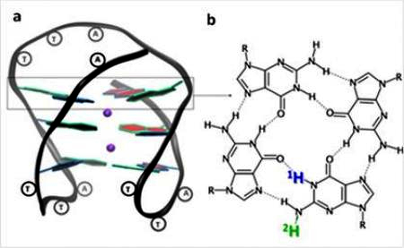
Figure 1: G-quadruplex of a DNA helix, formed by the human telomeric sequence GGGTTAGGGTTAGGGTTAGGG; the guanines (G) associate with each other by hydrogen bonds forming tetrads (b). After photoionization, the loss of a proton (blue or green) leads to the formation of two different deprotonated radicals.
In order to better understand the oxidative damage caused by UV radiation on DNA telomeres, the researchers undertook a quantitative study of emitted electrons and radicals formed during irradiation. For this, they used time-resolved absorption spectroscopy, which allows transient radical experiments to be identified by their absorption spectrum, coupled with quantum chemistry calculations. The human telomeric sequence, capable of folding into four-stranded structures called G-quadruplexes, has been more particularly studied. The quantum yield observed for one-photon ionization at 266 nm (4.66 eV) is 4.5 x 10
-3, comparable to that of other phototoreactions such as dimerization of bases, well known as a source of damage to the DNA under these conditions.
Following the ejection of electrons, the transient radicals, observable within the G-quadruplex, are identified by their absorption spectrum. The radical cation initially formed can either directly react chemically or simply lose a proton. This loss makes the radical electrically neutral, before it reacts in other ways. Thus, in less than 2 μs, 50% of the initial cation radicals lose their proton which is not engaged in a hydrogen bond (in green in Figure 2). These radicals disappear in less than 5 ms. Unexpectedly, another deprotonation pathway of the cation radical is observed, with the loss of the blue inner proton, which occurs in 5 ms. The life of this neutral radical is much longer, of the order of 50 ms.
Figure 2: Evolution of the population (in %) of the different radicals identified: (G) +.: cation radical - (G-H2).: deprotonated radical, with loss of the external hydrogen atom - (G-H1).: deprotonated radical, with loss of the internal hydrogen atom
Several important conclusions have been drawn from this work:
- The secondary structure plays a key role for the phenomenon of photoionization which is detected only for G-quadruplexes and not for the single-stranded telomeric sequence.
- Three types of formed radicals have been identified: the radical cation and two deprotonated radicals, having lost the external proton or the inner proton; the corresponding chemical reactions remain to be discovered.
- Radical cations, which are the charge carriers (electron-holes), survive in G-quadruplexes 1000 times longer than in single and double DNA helices
[3], [4]. This point may prove useful in developing bio-inspired nanotechnology devices.