 Background: Promises and challenges of the Si anodes
The recent rapid increase of the demand of higher energy density along with higher power density lithium-ion batteries (LiBs) requires the development of advanced cathode/anode materials with higher capacity. Among which, silicon-based anode have drawn particular interests due to its very high gravimetric capacity, ten times higher than graphite materials (3579 vs 371 mAh g-1)[1]. However, the resulting high volumetric variation upon lithiation/delithiation leading to poor cyclelife and associated important lithium inventory loss, have impeded its large scale development. These challenges can be addressed providing that powerful characterization tools are able to probe the degradation phenomena occurring at multiple scale during the electrode cyclelife. Among these, X-ray tomography has been used as non-invasive 3D investigation tool in order to probe at different length scale the electrode microstructure. Moreover, phase contrast imaging has brought to light a practical way to enhance visibility between weak absorbing materials and/or small details of differing refractive index within structure, and thus accessing a sharpen overview of the 3D morphology of complex material, which is of particular interest in the frame of energy-related materials. This is particularly relevant when accessing large scale facility (ESRF), which offer very high brightness and coherency allowing very high spatial resolution and enhanced contrast between low attenuating materials.
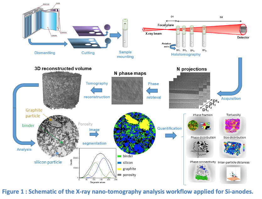
Electrode microstructure: X-ray nano-tomographyThe X-ray tomography is based on the recording of attenuation images on a CCD/CMOS camera along the rotation of a sample exposed through the X-ray beam (synchrotron or laboratory). Combining these images in the reciprocal Fourier space allow reconstructing a 3D image of the sample volume based on the imaginary part of the refractive index of the materials, i.e. attenuation-based. In order to increase the sensitivity towards low attenuating materials and provide enhanced contrast, phase contrast tomography is of outmost interest for accessing the real part of the refractive index. In particular, at the ID16B beamline [2] at the ESRF, the holotomography technique [3] has been developed through the combination of multiple sample-detector distances for retrieving complementary phase information out of the interference patterns produced on the detector. Qualitative information can be directly interpreted from the 3D volume reconstructions, but most importantly quantitative analyses can be extracted provided a thoroughly segmentation step is performed before. Subsequently, a large panel of parameters can be measured, such as the volume fraction, size distribution, intra and inter-connectivity, geometrical tortuosity, etc. for the different labeled phases (Fig. 1). In order to label or segment a phase, different approaches can be investigated, either simply based on the histogram deconvolution through Otsu thresholding or more complex using machine learning based algorithm, e.g. random forest.
 The internship
On key aspect of the degradation is the study of the SEI formation and its evolution along the cyclelife of the electrode depending on operation conditions (Fig. 2).
 In this context, the aim of the intern is to analyze 3D X-ray tomography images obtained on silicon-graphite composite electrodes, aged using different electrolyte composition (standard vs highly concentrated) and under different temperature conditions (room temperature and 40°C). Acquisitions have been performed at ESRF on post mortem samples at the lithiated or delithiated states for different steps of their cyclelife. The main task will be the processing and quantitative analysis of the large amount of tomography images in order to highlight the key factor influencing the SEI cumulative build-up. Participation to experimental campaigns of measurements at the synchrotron will be possible depending on the schedule of the runs in the internship period. The analyzed results will be presented and shared across a consortium of European academic laboratory, with possible collaboration with modelling teams. The intern will be working for 6 months at CEA in Grenoble, a major French National Lab focused on the development of carbon-free innovative solutions.
The candidate will benefit from an interdisciplinary research environment at the cutting edge of chemistry, physics and nanoscience. He/she will be trained in the synthesis of semiconductor quantum dots and their optical/structural characterization, and will get insight in the application of these nanoparticles in biological applications (e.g.in-vivo imaging, bio-sensing)
Requested skills
M2 master student with knowledge in X-ray tomography and image analysis (Fiji, Ilastik, Python) and/or energy storage devices (especially Si-anodes and Li-ion batteries) will be important.
Information
Starting date: February - March 2024
Duration: 5- 6 months
Laboratory / Team : CEA LITEN/SAMA/LMP & CEA-IRIG. SyMMES / STEP
(https://www.symmes.fr/STEP & https://liten.cea.fr/cea-tech/liten/Pages/Accueil.aspx)
Keywords: Li-ion batteries ; silicon anodes ; SEI ; synchrotron ; X-ray tomography imaging
This Master internship could be followed into a PhD within the same research area: eventually, depending on funding opportunities)